Access the webinar recording here

Questions regarding other body composition assessment tools:
- Q: Any informative ideas about 3D body scanning, similar to InBody Scan?
- Q: DXA is the gold standard test for BMD? Is it right?
- Q: What are your thoughts/opinions on using ultrasound in assessing body fat as compared to using DXA?
- Q: You mentioned BIA — what about the COSMED air-displacement assessments?
- Q: Any specific model to impedance measurement?
- Q: If you do not have a DXA, do you see utility in BIA measurements?
- Q: Do you have a favorite method of testing body fat percentage?
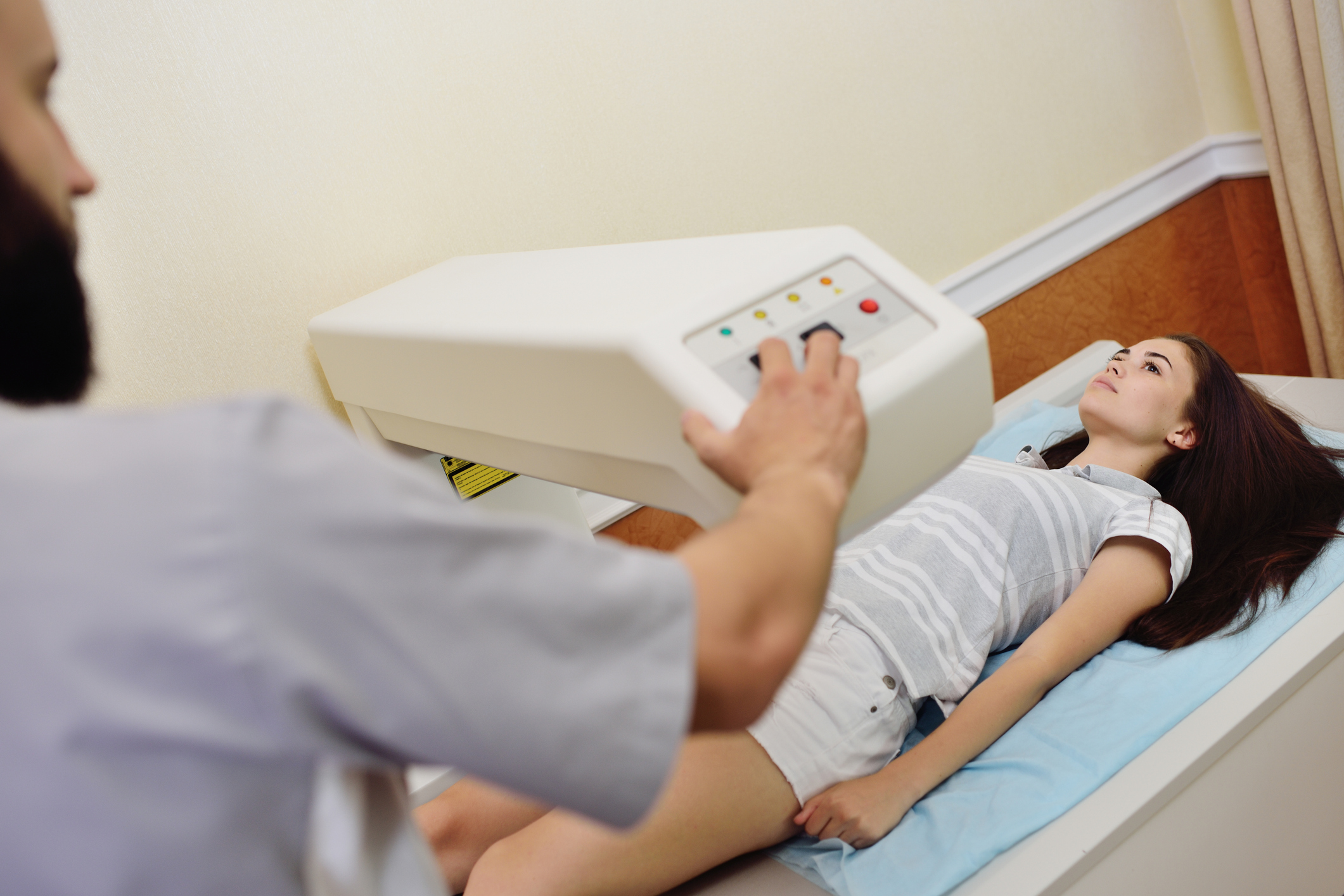
There are many pros and cons to any body composition assessment tool, DXA included. Many of you asked my thoughts about other methods — specifically bioelectric impedance analysis (BIA — InBody), air displacement (BOD POD), 3D scanners such as the SizeStream, and ultrasound — compared to DXA. DXA’s precision is greater than any of these measures and is the gold-standard tool for bone mineral density. So if you can afford one, I say go for it. But they are expensive, require radiation and special training, and are not mobile in most cases.
- BIA’s precision can be hydration dependent, but the newer InBodys provide a range for total body water, so as long as you’re scanning within that range, it is a good option that is cheaper and often portable.
- The BOD POD is useful but has size limitations and is also expensive. And if your client is having a hard time getting the breathing technique down, the standardized equations for residual volume are not great in my opinion and can increase error rates.
- 3D scanners are an interesting newer player to the field. With no radiation requirements, size limitations or hydration issues, they provide a quick option. Be sure to pay special attention to which regression equation you use in conjunction with the anthropometrics reported; they vary greatly. If you’re interested, please read Body Fat Assessment Techniques: Is There a Place for 3D Body Scanners? for more information about the validity of this tool.
- Lastly, the use of ultrasound to estimate body fat percentage: Ultrasound provides tissue thicknesses and estimates of muscle quality (via density) and is a useful tool for any research team. However, measuring subcutaneous adipose tissue in one or two areas will not be a great tool for the estimate of total body fat. It can, however, be used to assess site-specific fat loss over time.
In summary, my favorite body composition tool is DXA, but that is because I am in a research setting that helps me circumvent its limitations. If you’re in the field and need something cheaper and more portable but don’t want to go the skinfold/circumference tape route, I would recommend a newer InBody from the list above. 
Questions regarding scanning procedures:
- Q: Any observations in dealing with tactical athletes or older athletes with joint replacements?
- Q: Do you have any recommendations/best practices for using Custom RoI while performing segment-wise body composition?
- Q: Is it possible to mirror estimate data from left side to right side?
- Q: Do they irradiate the skull and gonads? If so, is this necessary to calculate a body composition evaluation?
- Q: Hi, is there a safe frequency for DXA scan? My lecturer used to say limit to four scans/year due to radiation.
- Q: What are your thoughts on scanning tall athletes as total body less head (TBLH) to monitor longitudinally?
- Q: Does the temperature/humidity in the room influence the test results?
Lots of good questions fell within the topic area of scanning procedures. Folks had questions about working with tall athletes, tactical athletes, those with joint replacements, how to create novel scanning procedures, scan frequency, radiation exposures and room conditions for scans.
- When scanning anyone with orthopedic implants, the implant, regardless of synthetic material, will most likely inflate your site-specific BMC and total body aBMD Z- or T-scores. It doesn’t mean you can’t scan them. Instead, it will be more important to monitor the rates of change, as we assume the implant itself will not change from scan to scan. We often scan older adults with one or more arthroplasties, and with an inaccurate total body T-score, it is important to consider a site-specific scan (e.g., lumbar spine, hip, radius) to get a better idea of bone density measures from a clinical standpoint.
- Tall and wide athletes have special scanning procedures, and as noted in the talk, it is really important that you “have a plan before you scan.” For wide individuals, 95% of the time we just take the mirrored data from the right arm or leg and use it for the left arm or leg, which nearly all DXAs will automatically provide for you on the output. For tall athletes, many procedures exist, but most have the same limitation. The key consideration is to ensure the exact same parts of the body are scanned each time to be sure the changes you see over time are indeed real and not artifacts from different positioning from scan to scan. Some folks consider cutting off the head as an option, and although the density in the skull is less likely to change drastically, many tall athletes will require their head and parts of the foot to be outside the scan area. The technique we proposed is to create a region of interest (ROI) that can be placed onto the head and will never change length so, assuming your athlete is no longer getting taller, you can be sure the same exact tissue is being scanned each time.
- The DXA scan uses radiation to scan the entire body, so yes, that will include the skull and gonads, and their inclusion is part of the total body scan for the assessment of body composition. There is no way to exclude these regions from a total body scan and still get “total body fat percentage.”
- The timing throughout the year and frequency of the scans are up to you and your team, but most of the time, scanning more than four times per year will not exceed the machine’s error rate and should not be reported. The timing of the year may change the ambient environment of the room in which the DXA is housed (e.g., summer is hot, winter is cold). It is important that your machine is housed within a room that is level, very clean and maintains a normal temperature. I would not recommend having a DXA in a space that lacks climate controls. Before you get a DXA delivered, most companies will perform a site visit to ensure the space is suitable for the machine’s successful long-term performance.
Questions regarding data reporting and management:
- Q: When working with younger athletes (between 16 and 18 years) do you use the Z-score based on length or the Z-score based on age? Since we get both those Z-scores when we scan younger athletes.
- Q: I appreciate Dr. Baker mentioning the issues with DXA and the Oregon track program. Could she speak more about her experience with reporting and using data/images among athletes/coaches that discourages unhealthy use of the data?
- Q: I did not manage to register the web address with the automated DXA-result extraction … is there any place or in any way we could get access to this?
- Q: Hi Dr. Baker, do you compare to the NHANES database with the scans at your practice?
- Q: I agree with T-score reporting for those over 50 years. However, can you explain to all as to why?
- Q: Can you recommend a source for the most current guidelines on bone health for general populations?
- Q: Do you think results may differ between Caucasians and Asians?
- Q: How do you educate people that have some curvature in their spine over multiple scans and express anxiety or distress about that?
- Q: Can DXA provide us with details about scoliosis?
- Q: Could a difference in bone density between two legs also correlate with a difference in muscle mass?
- Q: What is your role within the sports teams you work with? How do you organize meeting with the coaching staff?
- Q: Did you say there was an app for DXA data?
- Q: Would it be a good idea to use DXA for clients who have bone issues like arthritis? Would that be within the scope of practice for a personal trainer?
Many of you submitted great data-extraction, management and reporting questions. Below are my thoughts on the most common.
- First: Clinically, Z-scores are biological sex-, age- and ethnicity-matched data that are to be used for those 50 and under (including those under the age of 20). Different ethnicities have greater or lower aBMD, so that is an important factor to consider. T-scores are the clinical tool for those over 50 years and are compared to a 30-year-old reference sample. This doesn’t mean you cannot discuss Z-scores with those over 50 years; in fact, it often helps them to put into perspective where they are. But the T-score is what needs to be used for clinical diagnosis. Your DXA will have specific databases it uses to create these scores, which are often in part of NHANES. The bone mass will be strongly correlated with lean mass and total body mass; this statistical relationship also holds true for most site-specific locations, such as increased bone in the leg with increased muscle.
Those with osteoarthritis often have site-specific increases in bone mineral density as the damaged bone becomes sclerotic. For instance, someone with osteoarthritis in their lumbar spine will have an inflated aBMD and T-score, so instead we need to perform a scan of the hip or radius to get more accurate information regarding aBMD clinical classifications.
Lastly, spinal deviations such as scoliosis are often evident on total body scans. The deviation may be in part due to poor positioning, muscle imbalances, or actual skeletal deviations. But unless you are a medical doctor, most states will not allow you to make a diagnosis. Encourage the subject to share their scans with their primary care physician. This last point is true for any clinical diagnosis you see: Educate them and encourage them to see their doctor. You can provide bone recommendations such as good dietary habits (e.g., calcium and vitamin D), exercise that is weight-bearing, and getting a DXA if someone is concerned — but again, be careful here. Most states do not allow you to make recommendations for bone health if you don’t hold an M.D. or D.O.
- The DXA provides an immense amount of data beyond the overused and abused body fat percent, but getting these data can be cumbersome and result in transcription errors. As noted in the talk, please consider automating this process. There are many ways to do this, but we use the steps detailed in the following paper: DXA2: An Automated Program for Extraction of Dual-Energy X-Ray Absorptiometry Data.
- Lastly, data reporting is the responsibility of the person who runs the DXA. In my case, any scans of athletes (regardless of who did the scan), are my responsibility. In our experience, the athletes want to learn about their bodies and are inherently curious, but very few people can see their images or get their BF% number and have a good response. We do not and will not provide athletes their BF% or the body composition number; instead, we take the time to educate them about their muscle and bones and how those two tissues are vital for performance and injury prevention. This procedure is something that is clearly discussed with the coaching staff well before we ever begin working together. My goal is to work in conjunction with all of the team’s support staff, not in opposition. Different entities want different data at different times, and we work to provide those data to them in a way they can easily understand and then use. My role within a team setting will be unique to each team and the relationship that has been cultivated with the coaching staff. I do not think a one-size-fits-all approach best serves the athletes or the coaches.
Questions regarding precision testing:
- Q: I work in corporate health and wellness, so this is out of my field. But I want to ask about the error percentage. When you do a percentage test you get the error percentage, but how would you use that when gathering your data? Do you subtract that or add the % error to the data, or am I completely off?
- Q: The least significant change and the smallest worthwhile change — are they the same?
- Q: Hologic and GE provide default LSC values.
- Q: I’m thinking of the CV/LSC and wonder how you interpret repeated measurements. And the changes you detect: Do you first subtract the variation and then register the change, or do you register the exact measured change?
- Q: Can default LSC values for the DXA be used?
- Q: How often do you do precision testing?
Answers:
- It is ok if you have not completed a precision test of your machine yet, but if you plan on using the data to make inferences about body composition changes over time, it is 100% necessary. Two important things to consider is that the precision, or the machine’s error, is a function of both the machine and your scanning techniques. So it is not appropriate to just use the manufacturer’s cited precision values. So, when the machine undergoes major repair or someone new in the facility becomes the primary technician, it is important to repeat the test. First read the International Society for Clinical Densitometry’s recommendations. You can scan 15 folks three times or 30 folks two times and then plug in the BMC, aBMD, lean mass and fat mass data into their calculator to determine the precision of your machine.
- The second really good question I have gotten is how do I report my data in light of the precision test’s results? As a researcher, I run statistical tests to understand if changes over time were due to chance or the intervention, so when my significance is p < 0.05, we consider it statistically significant. But despite the p value, if the measure does not exceed the least significant change absolute value or the coefficient of variation (CV%) then those data are most likely not clinically significant and need to be reported as such.